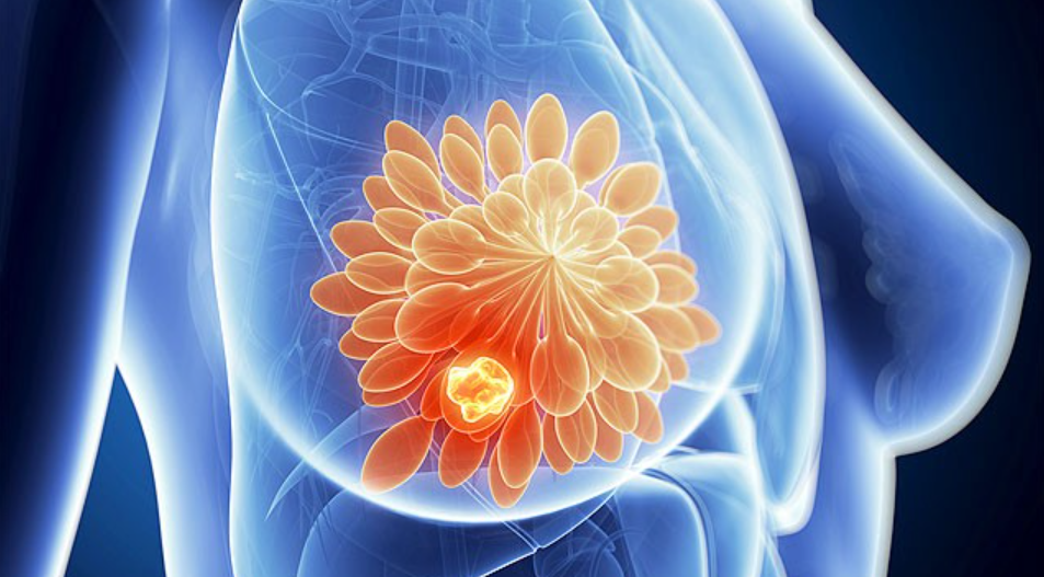
BREASTS; EVERYONE HAS THEM. DO YOU KNOW HOW THEY WORK?
Understanding the mystery of the mammary glands.
We all have breasts. Structurally, breasts are simply modified sweat glands. But functionally, the mammary glands produce milk, life- saving nutrition for newborn babies allowing the human race to evolve and flourish.
The mammary glands are wondrous and complex structures. Let’s take a tour of the breast anatomy.
What are the mammary glands?

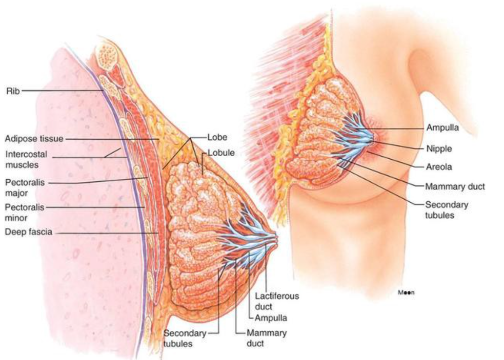
Used with permission from Medika Life
Mammary glands are located in the breast overlying the pectoralis major muscles. Both males and females have them, but they are functional only in women.
Externally, each breast has a raised nipple surrounded by a circular pigmented area called the areola. The nipples are sensitive to touch because of a dense array of nerve fibers and smooth muscle that contracts and causes them to become erect in response to stimulation.
Internally, the adult female breast contains 15 to 20 lobes of glandular tissue that radiate around the nipple. The lobes are separated by firm connective tissue and fatty adipose tissue.
The connective tissue helps support the breast. Bands of connective tissue, called suspensory (Cooper’s) ligaments, extend through the breast from the skin to the underlying muscles. The amount and distribution of the adipose tissue determine the size and shape of the breast. To keep it simple, fatty tissue determines breast size and connective tissue holds them up.
A complex plumbing system
Have you ever wondered how milk gets to the nipple? Each lobe consists of lobules that contain the milk-producing glandular units. A tube called a lactiferous duct collects milk from the lobules and carries it to the nipple.
Milk is stored in a reservoir called the ampulla (lactiferous sinus) just before arrival at the nipple. After the sinus, the duct splits and narrows allowing multiple ducts to bring milk to the surface of the nipple.
Mammary gland function is regulated by hormones
Four hormones have an integral role in breast development. Estrogen levels increase during puberty and stimulate the production of glandular breast tissue. Estrogen increases adipose tissue causing the breast to increase in size. Progesterone plays a role as well by stimulating the development of the duct system.
During pregnancy, these hormones further enhance the development of the mammary glands. Prolactin from the anterior pituitary gland stimulates the milk production inside of the glandular tissue, and oxytocin triggers the ejection of milk from the glands.

Used with permission from Medika Life
Breast surface anatomy
The breast is located on the anterior thoracic wall. It extends horizontally from the lateral border of the sternum to the mid-axillary line. Vertically, it spans between the 2nd and 6thintercostal cartilages. It lies superficially to the pectoralis major and serratus anterior muscles.
The breast is composed of two regions. The circular body is the prominent area of the breast. The axillary tail is a smaller area running along the inferior lateral edge of the pectoralis major towards the axillary fossa.
At the center of the breast is the nipple. It is made mostly of smooth muscle fibers. The pigmented area of skin surrounding the nipple is called the areolae. There are numerous sebaceous glands within the areolae . These glands enlarge during pregnancy, secreting an oily protective lubricant for the nipple.
The breast’s blood supply
The middle of the breast receives blood from the internal mammary artery, a branch of the subclavian artery.
The lateral part of the breast receives blood from four vessels:
- Lateral thoracic and thoracoacromial branches — originate from the axillary artery.
- Lateral mammary branches — originate from the posterior intercostal arteries (derived from the aorta). They supply the lateral aspect of the breast in the 2nd 3rd and 4th intercostal spaces.
- Mammary branch — originates from the anterior intercostal artery.
The breast lymphatic system

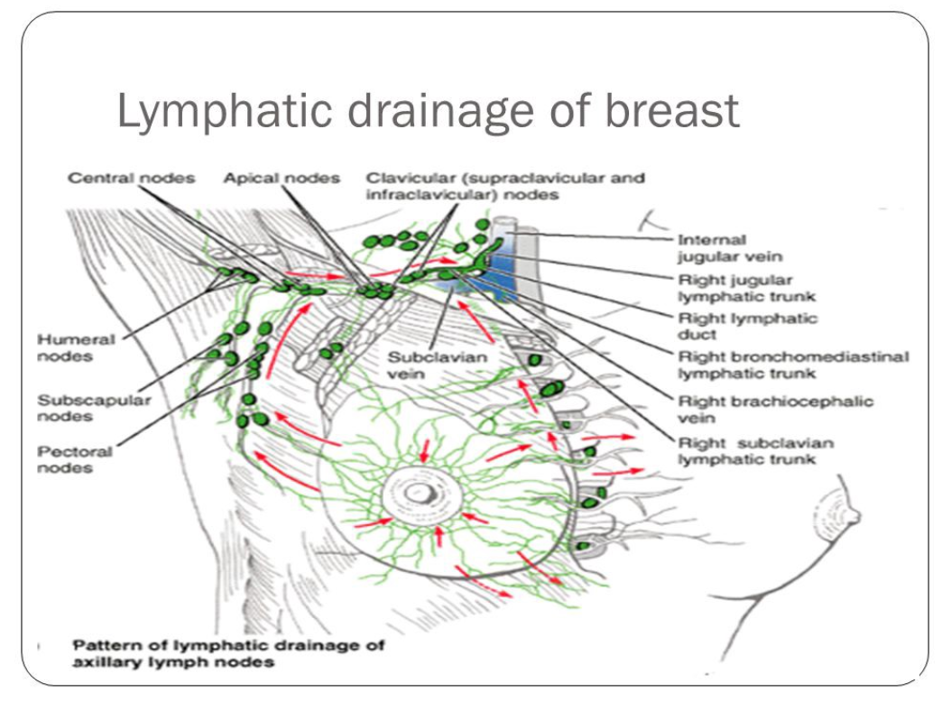
Used with permission from Medika Life
The lymphatic drainage of the breast is incredibly important. Breast cancer spreads through the lymph when it spreads (metastisizes).
There are three groups of lymph nodes that receive lymph from breast tissue — the axillary nodes (75%), parasternal nodes (20%), and posterior intercostal nodes (5%).
Thank you to BeingWell for publishing this article on Medium.
Originally posted on Medika Life
Blog Author: Dr. Richard Wagner
Main Blog Photo By: Jan Kopřiva on Unsplash

